
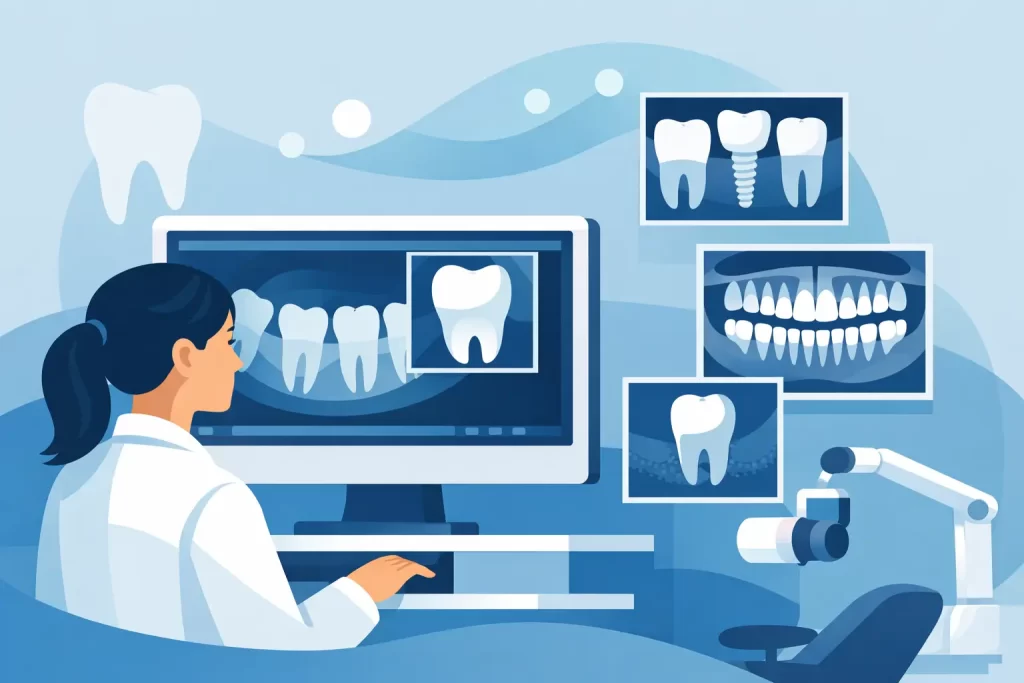
What Digital X Rays at the Dentist Can Show
A tooth can look perfectly fine in the mirror and still have a problem developing beneath the surface. When you search for a “digital x rays dentist,” you are likely looking for a practice that can see beyond what is visible, explain what is happening clearly, and help you make decisions before a small concern becomes a painful emergency.
At Restorative Dental Jamaica, digital X-rays are one part of a careful, patient-centered examination. They help our team assess teeth, roots, bone, and areas between teeth that cannot be evaluated fully with the naked eye. The goal is not to take images unnecessarily. It is to gather the right information for thoughtful care that protects your natural smile whenever possible.
Why Digital X Rays at the Dentist Matter
A dental examination begins with listening to your concerns, looking closely at your teeth and gums, and reviewing your oral health history. However, some of the most common dental problems begin in places a visual exam cannot reach. Decay can form between teeth, an old filling can leak around its edges, or an infection can develop near the tip of a tooth root.
Digital X-rays give your dentist a more complete view. The images appear quickly on a computer screen, allowing us to review them with you during your visit. Rather than hearing that a tooth “needs work” without context, you can see the area of concern and understand why a filling, crown, root canal treatment, or monitoring plan may be recommended.
This clarity is especially valuable when a tooth is not causing pain. Many people assume that no discomfort means no dental problem, but early decay and gum-related bone loss can be silent. Finding concerns early often means simpler treatment, less time in the dental chair, and a better chance of preserving the tooth.
What a Digital Dental X-Ray Can Reveal
Different X-ray views are used for different clinical questions. Bitewing images are commonly used to check for cavities between back teeth and to assess the condition of existing fillings or crowns. Periapical images show an entire tooth, including its root and the bone surrounding it. This can be helpful when investigating tooth pain, sensitivity, swelling, or a possible infection.
A panoramic X-ray provides a wider view of the jaws, teeth, and surrounding structures. It may be recommended when planning extractions, assessing wisdom teeth, considering dentures, or evaluating certain orthodontic concerns. Your dentist will select the type of image based on your needs rather than using the same approach for every appointment.
Digital images may help identify:
- Cavities between teeth or beneath older restorations
- Infection or inflammation around a tooth root
- Bone changes associated with gum disease
- Cracks, trauma, or changes affecting a painful tooth
- Impacted or poorly positioned teeth, including wisdom teeth
- Areas that need attention before crowns, dentures, aligners, or other treatment
An X-ray is a diagnostic tool, not a treatment decision by itself. Your dentist considers the image alongside your symptoms, exam findings, dental history, and personal goals. A shadow on an image may call for treatment, closer monitoring, or sometimes no action at all. The right choice depends on what the image shows and how that finding relates to your overall oral health.
Digital X Rays Dentist Patients Can Feel Comfortable With
Many patients remember older dental X-rays as awkward, slow, or uncomfortable. Digital technology improves much of that experience. The image is captured electronically, so there is no film to develop and less waiting for results. Your dentist can enlarge the image, adjust contrast when appropriate, and point out specific areas in a way that is easier to understand.
The process itself is usually quick. A small sensor or image receptor is positioned in the mouth, and you will be asked to hold still for a few seconds while the image is taken. Some views can feel a little awkward, particularly if you have a sensitive gag reflex or a sore area in your mouth. Let the dental team know before starting. We can work patiently, explain each step, and make adjustments where possible to help you stay comfortable.
Patients also understandably ask about radiation. Dental X-rays use a low dose of radiation, and digital systems typically require less exposure than traditional film X-rays. Protective measures and careful imaging techniques are used to keep exposure as low as reasonably achievable. X-rays are taken only when they can provide useful information for your care.
If you are pregnant, think you may be pregnant, or have questions about your medical history, tell your dentist before your appointment. Dental care can often still be planned safely, but the timing and type of imaging should be considered individually. Open communication helps your care team make the most appropriate recommendation.
How Often Are Dental X-Rays Needed?
There is no single schedule that fits every patient. The frequency of X-rays depends on your risk of tooth decay, gum health, age, existing dental work, symptoms, and how long it has been since your last images. A patient with a history of frequent cavities may need bitewing X-rays more often than someone with stable oral health and low cavity risk.
Children and teenagers may also need images at different intervals because their teeth and jaws are changing. For adults, images may be advised when there is pain, a broken tooth, swelling, a loose crown, a suspected infection, or a treatment plan that requires a detailed view below the gumline.
If you have recent X-rays from another dental office, bring them or arrange for them to be shared when possible. This may reduce the need to repeat images. Still, if the available images are outdated, incomplete, or do not show the area causing concern, new X-rays may be necessary for an accurate diagnosis.
Better Planning for Restorative and Preventive Care
Digital X-rays are particularly useful when planning care designed to repair and preserve teeth. Before placing a crown, your dentist may need to check the tooth root, surrounding bone, and the extent of decay. Before root canal therapy, images help evaluate the infection and guide treatment. When discussing dentures or an extraction, X-rays can reveal important details about tooth roots and jawbone structure.
They also support preventive care. A small cavity found early may be treated with a conservative white filling. If decay reaches deeper into the tooth, treatment may become more complex and costly. Seeing changes sooner gives you more options and more control over your dental health.
For cosmetic treatments, X-rays do not determine whether a smile will look beautiful, but they help confirm that the foundation is healthy. Whitening, veneers, bonding, and aligners are best considered after your dentist has checked for hidden decay, infection, or gum concerns that should be addressed first.
What to Expect at Your Appointment
If digital X-rays are recommended, your dental team should explain why they are needed and what information they may provide. You can ask questions at any point, including whether a different image is needed, how recent images may be used, or what the findings mean for your treatment choices.
After the images are taken, your dentist can review them with you and discuss the next step in plain language. That may be routine monitoring, preventive treatment, a filling, a crown, root canal therapy, or a referral when specialized care is appropriate. You should leave knowing what was found, why a recommendation was made, and what options are available.
A clear image can turn uncertainty into a practical plan. If you have tooth pain, have not had a dental assessment in some time, or simply want confidence that hidden problems are not being missed, scheduling an examination is a caring step toward protecting the smile you use every day.
Leave a reply








Leave a reply